ADAS 3D
Overview
ADAS 3D is a medical software tool developed by Adas3D Medical S.L, specifically designed for electrophysiologists. It specializes in the post-processing of cardiovascular enhanced Magnetic Resonance (MR) images and Computed Tomography Angiography (CTA) images, which are formatted in the Digital Imaging and Communication in Medicine (DICOM) standard.
The software aids in the non-invasive calculation, quantification, and visualization of 3D cardiac imaging data, supporting a comprehensive diagnostic decision-making process for understanding cardiovascular disease. It is particularly useful for patients with myocardial scar produced by ischemic or non-ischemic heart disease.
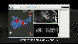
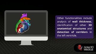
ADAS 3D consists of two main modules: ADAS 3D LV (Left Ventricle) and ADAS 3D LA (Left Atrium). These modules automatically identify areas of fibrosis in the myocardium and create detailed 3D models of the patient’s anatomy, including adjacent heart structures. The tool helps in visualizing fibrosis, wall thickness, and surrounding anatomical structures, enabling electrophysiologists to extract key clinical information for decision-making, planning, and guidance of electrophysiology (EP) procedures.
While the software is intended to assist healthcare professionals in diagnosis, it is not meant to fully substitute their clinical judgment. It is available through platforms like the Siemens Healthineers’ Digital Marketplace, integrating with syngo.via OpenApps.
Reviewed by Pouyan Golshani, MD — Interventional Radiologist
Key Features
- Post-processing of cardiovascular enhanced MRI and CTA images
- Calculation, quantification, and visualization of 3D cardiac imaging data
- Identification of fibrotic tissue in the left ventricle (ADAS 3D LV)
- Identification of fibrotic substrate in the left atrium (ADAS 3D LA)
- Automated segmentation for specific image sequences (AI-powered, coming soon)
- Export of 3D models to electrophysiology (EP) navigation systems
- Analysis from LV Enhancement: Visualization in concentric layers, detection of anatomical corridors, identification of 3D core tissue
- Tissue characterization with automatic classification based on Image Intensity Ratio
- Provides a roadmap to assist in EP procedures
- DICOM image database processing
Use Cases
- Planning electrophysiology interventions
- Guidance during electrophysiology procedures
- Identification of arrhythmia substrate
- Visualization and analysis of cardiac MR and CTA images for cardiovascular disease
- Understanding fibrosis distribution in the myocardium
- Pre-procedural and peri-procedural clinical decision making
What Physicians Need to Know
ADAS 3D is a valuable pre-procedural planning and intra-procedural guidance tool for electrophysiologists. Leverage its capabilities to quantify myocardial fibrosis and visualize complex 3D cardiac anatomy from MRI and CT images, which can significantly enhance the understanding of arrhythmia substrates for ablation procedures. Remember that the software is intended to assist healthcare professionals and should complement, not substitute, clinical judgment. Pay attention to the quality and resolution of input DICOM images, as they directly impact the accuracy of the generated data. Explore the upcoming AI-powered automated segmentation for potential workflow efficiencies.
ADAS 3D supports importing cardiac images in DICOM format and can export its generated 3D models and fibrosis information to various electrophysiology (EP) navigation systems. It is available in a Cvi42 integrated version and is integrated into Siemens Healthineers' syngo.via OpenApps via their Digital Marketplace. The software has also been integrated with Stereotaxis robotic magnetic navigation technologies for enhanced procedural guidance.
Details
| Category | Cardiology AI, Radiology & Imaging AI, Surgical AI |
| Pricing | Contact for pricing — Not disclosed |
| Deployment | Stand-alone software tool, can be integrated with imaging systems (e.g., Siemens Healthineers' Digital Marketplace). |
| Compliance | |
| BAA Available | Unknown AI-estimated |
| HIPAA Compliant | Unknown AI-estimated |
| FDA Status |
1 AI-estimated ADAS 3D has received multiple FDA 510(k) clearances (K191125, K210850, K212421, K230803, K240791) and CE Mark certification. It is indicated for use in clinical settings to support the visualization and analysis of MR and CT images of the heart for patients with cardiovascular disease, specifically those with myocardial scar. |
| Integrations | |
| EHR | Not specified |
| Specialties | Cardiology, Radiology |
What the Web Says
ADAS 3D, developed by Adas3D Medical S.L., is a software designed for the visualization and analysis of enhancement in MR and CT cardiovascular images for patients with cardiovascular disease, particularly those with myocardial scar. It helps physicians identify potential sources of irregular heartbeats and provides 3D cardiac models to guide treatment, especially for ventricular tachycardia and atrial fibrillation ablation procedures. The software integrates with robotic magnetic navigation technologies, allowing physicians to use the generated images to guide therapy. While the clinical significance for identifying arrhythmia substrates for treatment or risk stratification has not been fully established, it is intended to support qualified medical professionals in clinical decision-making.
Overall: PositiveStrengths
- Supports rapid and precise identification of problematic cardiac tissue.
- Generates detailed three-dimensional cardiac models from preoperative cardiac MRI.
- Can be used to define the shape and fibrosis distribution of the Left Atrium for atrial fibrillation procedures.
- Integrates with robotic magnetic navigation technologies for guided therapy during cardiac ablation.
- High concordance with electroanatomical mapping for myocardial tissue characterization.
- Provides visualization and analysis of MR and CT images of the heart for cardiovascular disease.
Limitations
- The data produced by ADAS 3D should not be used as an irrefutable basis for clinical diagnosis or patient treatment.
- The clinical significance of using ADAS 3D to identify arrhythmia substrates for treatment or risk stratification has not been established.
- Accuracy is dependent on the quality and resolution of the medical images processed.
- Limited public reviews available from independent sources like Reddit, G2, or Capterra specifically for ADAS 3D Medical's product.
Based on reviews from: Cardiac Rhythm News, PubMed Central (PMC), accessdata.fda.gov, Siemens Healthineers Digital Marketplace
Last updated: 2026-07-17
Ratings & Reviews
No reviews yet. Be the first to review this tool!
Rate ADAS 3D
Press & Coverage
Videos
Product demos, reviews, and walkthroughs for ADAS 3D.
ADAS 3D for the Left Ventricle