breastscape

by Olea Medical  · Based in France → — Improved Diagnosis for Life
Oncology Radiology

Contact vendor
Regulatory Status Disclosed

Overview

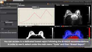
Olea Medical’s breastscape® V1.0 is an advanced, multi-vendor post-processing software solution designed to enhance and streamline breast MRI examinations. It supports radiologists and MRI technologists in the analysis, interpretation, and follow-up of MRI examinations, aiding in crucial daily decision-making. The solution incorporates cutting-edge AI algorithms and deep learning tools, tested by clinical experts, researchers, and imaging specialists, to overcome the limitations of traditional medical imaging. breastscape® offers semi-automatic lesion diagnosis, automatic lesion detection, and integrates BI-RADS® reporting, significantly improving diagnostic capabilities and patient care. It also includes a dedicated module, BreastLoc, for MRI-guided breast biopsy planning. The software provides quantification, volumetrics, tissue segmentation, and advanced visualization tools like 4D MIP, MPR, and automatic subtraction, along with motion correction for more confident image interpretation. Deployable on-premises or in the cloud, breastscape® is built to optimize clinical productivity and standardize workflows in the demanding field of breast imaging.

Reviewed by Pouyan Golshani, MD — Interventional Radiologist

Key Features

  • Semi-automatic lesion diagnosis in MRI
  • Automatic lesion detection
  • BI-RADS report integration
  • MRI Breast biopsy diagnostic tool (BreastLoc)
  • Quantification, volumetrics, and tissue segmentation
  • Motion correction
  • Longitudinal analysis
  • Multi-vendor post-processing solution
  • Advanced algorithms for characterization and follow-up of breast abnormal areas
  • 4D MIP, MPR, and automatic subtraction visualization tools

Use Cases

  • Breast lesion diagnosis in MRI
  • MRI Breast biopsy planning
  • Analysis, interpretation, and follow-up of breast MRI examinations
  • Screening women with dense breasts
  • Assessing evolution of suspected findings and response to treatments
  • Streamlining workflow for radiologists and MRI technologists

What Physicians Need to Know

Key Capabilities
Breastscape, part of Canon Medical's advanced visualization solutions, offers tools for breast cancer detection, characterization, and staging. It provides multi-parametric analysis, including MPR and 3D visualization, volume segmentation, multiple series fusion, kinetics, and curve analysis. The system also includes BI-RADS® ATLAS reporting to improve communication among radiologists, patients, and referring physicians. It supports automated 3D lesion volume and measurement computation, kinetic analysis, and lesion characterization. The AI-enabled BI-RADS® function on Aplio i-series can identify breast structures and suggest their size and category. The Aplio i-Series offers advanced breast imaging technologies such as 24 MHz ultra-high frequency linear transducers, Superb Microvascular Imaging (SMI) for vascularity assessment, BEAM (Biopsy Enhancement Auto Mode) for enhanced needle visualization, and MicroPure for visualizing micro-calcifications. Shear Wave Elastography (SWE) provides real-time visualization and numerical indicators of tissue stiffness.
Clinical Utility
Breastscape is designed to aid physicians in automating and simplifying MR breast screening and characterization, supporting confident and accurate diagnoses. It helps in evaluating dense breast tissue, performing adjunctive screening, and guiding interventional procedures. The tool aims to enhance diagnostic confidence and streamline workflows for radiologists, department managers, and sonographers. Early detection of breast cancer, especially when tumors are smaller than 1 cm, significantly increases survival rates. The system's ability to visualize fine details, micro-calcifications, and assess lesion stiffness contributes to more accurate diagnoses and treatment planning.
Integration Options
Breastscape is integrated into Vitrea Advanced Visualization, a multimodality, vendor-neutral solution that standardizes and consolidates radiology IT footprints. Canon Medical systems are designed to embrace open network standards, complying with DICOM and IHE standards for seamless integration into various network environments. The system can be used on-system with mammography images for direct comparison of suspicious areas. Reference Imaging features allow real-time comparison with mammography, MRIs, and previous ultrasounds. Canon's AI-integrated workflow connects hardware platforms with post-processing solutions like Vitrea and Olea, creating a streamlined pipeline from image acquisition to diagnostic review.
Compliance Status
The BreastApp module within Breastscape complies with BI-RADS® Atlas standardized breast imaging reporting guidelines. Canon Medical Systems' software development life cycle (SDLC) is governed by the NIST 800:53 Risk Management Framework, adhering to Defense Health Agency (DHA) certification requirements. Canon Medical Systems USA, Inc. maintains various conformance statements, including DICOM, IHE, HL7, and ISO certifications.
Pricing Model
Canon Medical offers flexible service agreements, including full security, in-house support, limited risk, and partnership options, allowing customization based on needs. Preventative maintenance agreements are also available. CanonONE is a strategic business alliance agreement for acquiring, servicing, and financing imaging equipment. Specific pricing details for Breastscape are not publicly available.
User Experience
The Aplio i-series offers a user-friendly workflow designed for efficiency and precision, allowing more time for scanning and less time for adjusting settings and annotations. Smart Body Mark improves diagnostic confidence and workflow by displaying scanned and unscanned breast regions and automatically creating annotations. The system aims to make medical imaging smoother and more streamlined for both technologists and patients through innovative and simple-to-use solutions. The user interface is designed for intuitive use, ensuring a straightforward exam workflow.
Support Quality
Canon Medical Systems USA provides 24/7 support through its US-based Customer Solutions Center. They aim for an onsite response time averaging less than 3 hours nationally, with 90% of parts shipping within four hours. Three engineers are assigned per system, and cybersecurity solutions are included with any service coverage. Canon Medical has received awards for its service quality in CT, MR, and RF, based on customer satisfaction surveys. They offer proactive monitoring and remote diagnostics to identify and address issues before they disrupt clinical schedules.
Implementation Complexity
Canon Medical's solutions are designed for seamless integration into existing workflows. The company provides training and education through the Canon Learning Center, offering online courses, webinars, and in-person instructor-led classes. Physician training programs are accredited and provide practical experience. Canon Medical also offers remote assistance for protocol changes, training, and scan assistance.
Evidence Base
Canon Medical Systems conducts research and collaborates with medical institutions. They publish white papers and presentations on the clinical utility of their technologies, such as MicroPure imaging in detecting breast calcifications and the clinical value of ultra-high frequency in breast ultrasound. Studies show that early detection of breast cancer significantly improves survival rates. Canon Medical also presents scientific papers at conferences like RSNA, detailing advancements in imaging techniques and their clinical relevance.
Physician Tip

Leverage the AI-enabled BI-RADS® function for automated structure identification and categorization to streamline your diagnostic workflow. Utilize the multi-parametric analysis capabilities, including kinetics and 3D visualization, for comprehensive lesion assessment. Take advantage of the on-system mammography image comparison and Reference Imaging features for real-time correlation with prior studies. Explore the benefits of Superb Microvascular Imaging (SMI) and Shear Wave Elastography (SWE) for detailed vascularity and tissue stiffness assessment, which can enhance diagnostic confidence.

Breastscape integrates seamlessly with Canon's Vitrea Advanced Visualization platform, providing a unified environment for multi-modality image review and analysis. The system adheres to DICOM and IHE standards, facilitating interoperability with existing PACS and other healthcare IT systems. This ensures a smooth workflow from image acquisition to reporting and archiving.

Details

Category Oncology AI, Radiology & Imaging AI
Pricing Contact vendor
DeploymentOn-premises, Cloud-based (AWS), Web-based
Compliance
BAA AvailableUnknown
HIPAA CompliantUnknown
FDA Status Cleared AI-estimated
Integrations
EHR Not specified
Specialties Oncology, Radiology

What the Web Says

Breastscape, developed by Olea Medical (a Canon Group Company), is a semi-automated software solution designed to assist physicians in the diagnosis, follow-up, and biopsy of breast lesions using MR imaging. It offers advanced visualization tools, automatic computation of parametric maps, and lesion segmentation, aiming to streamline the reporting process based on BI-RADS® Atlas. The software is intended for use by healthcare professionals and integrates with Canon Medical's Vitrea Advanced Visualization platform.

Overall: Positive

Strengths

  • Semi-automated solution for diagnostic assistance in breast lesions.
  • Provides automatic computation of parametric maps and automatic segmentation of lesions.
  • Assists with structured report preparation based on the latest BI-RADS® Atlas.
  • Offers advanced visualization tools like dynamic 3D MIP, MPR, and subtraction.
  • Enables lesion evolution follow-up and calculation of clinically relevant distances.
  • New report export format in PDF and improved report display.

Limitations

  • No specific cons were identified in the provided search results.
  • Limited information available from independent tech reviewers, Reddit, G2, or Capterra.
  • The product is a medical device reserved for health professionals, limiting broader public reviews.
  • The information largely comes from Canon Medical and Olea Medical, potentially lacking third-party critical analysis.
  • Specific details on integration with various healthcare IT systems beyond Vitrea AV are not extensively detailed.
  • While it assists, physicians must still review original images and interpret information.

Based on reviews from: medical.canon, healthaidb.com, slideshare.net, parikhradiology.com, scribd.com, olea-medical.com

Last updated: 2026-10-31

Ratings & Reviews

No reviews yet. Be the first to review this tool!

Rate breastscape

Clinical Value
Ease of Use
Integration
Support & Docs
Value for Money

Press & Coverage

usa.canon.com
MR Breast Advanced (Breastscape®)
MR Breast Advanced (Breastscape®) is a semi-automated solution for diagnostic assistance in breast lesions, providing automatic computation of parametric maps, lesion segmentation, and follow-up, with reports based on the BI-RADS® Atlas.
unknown
slideshare.net
Canon Medical Systems VISIONS Magazine - issue 31
This issue of Canon Medical Systems VISIONS Magazine, published in August 2018, features 'breastscape® v1.0 medical imaging post-processing software' as a medical device manufactured and marketed by Olea Medical®, a Canon Group Company.
2018-08
medical.canon
Vitrea Advanced Visualization 7.15 Release Notes
The Vitrea Advanced Visualization 7.15 release notes from January 2020 detail Breastscape® enhancements, including Lesion Merge, Early Signal Index, Advanced Breast extraction mask, and reporting features, along with easier modification to anatomical landmarks.
2020-01
medical.canon
Vitrea Advanced Visualization 7.16.0.SU02 Release Notes
The Vitrea Advanced Visualization 7.16.0.SU02 Release Notes mention Breastscape® 1.0 SP11 as part of the integrated software solutions, indicating ongoing updates and support for the product.
unknown
olea-medical.com
Breast MRI: an essential tool for early detection of breast cancer
This article from Olea Medical (a Canon Group Company) highlights the importance of breast MRI for early cancer detection and follow-up, emphasizing how specialized software solutions like Breastscape® streamline workflows and provide reliable results for radiologists.
unknown
olea-medical.com
Olea Medical's medical publications
Olea Medical, a Canon Group company, lists an article on the 'breastscape® v1.0' model, which precisely describes its automated functions such as exam registration, volume quantification, parametric maps, structured BI-RADS reports, and longitudinal follow-up.
2025-07
scribd.com
RSNA 2018 pages
Olea Medical, now part of Canon Medical Systems, promoted its new application dedicated to breast imaging, Breastscape v1.0, at RSNA 2018, which assists in characterization, follow-up, and planning of interventional breast procedures.
2019-01
nihs.go.jp
AI Medical Devices in Korea
A report on AI Medical Devices in Korea from August 2022 lists Breastscape v1.0 by Olea Medical as a Class 2a breast MR device, indicating its regulatory classification in the EU.
2022-08

Videos

Product demos, reviews, and walkthroughs for breastscape.

View all on YouTube

Frequently Asked Questions

Breastscape is designed as a complete breast MR platform that automates and simplifies breast screening, integrating with existing PACS systems for seamless workflow. It assists radiologists with visualization, analysis, and reporting for multiparametric breast MR exams, including automatic computation of parametric maps and lesion segmentation. Additionally, it offers tools for MR-guided breast biopsy planning, calculating lesion coordinates and needle insertion lengths.
Breastscape v1.0 is a medical device manufactured by Olea Medical, intended for use by breast imaging physicians and trained MRI technologists. It holds CE Certification (Class IIa) and FDA 510(k) clearance (Class II). As with any medical device, adherence to local regulations and guidelines for AI in healthcare is crucial, and patient management decisions should not be based solely on Breastscape's results.
While Breastscape offers advanced features, it is not intended for the primary interpretation of digital mammography images. Patient management decisions should always be made by a skilled physician and not solely based on the software's output. Additionally, the effectiveness of AI tools can be influenced by factors like data quality and the diversity of datasets used for training, which can impact generalizability.
Yes, the field of AI in breast oncology is evolving rapidly with various solutions. Canon Medical offers AI-enabled BI-RADS functions on its Aplio i-series ultrasound systems for breast structure identification and categorization. Other AI tools like IBM Watson for Oncology provide decision support for personalized cancer treatment, and PathAI assists in pathology slide analysis for cancer detection. Panoramic photoacoustic computed tomography (PACT) with machine learning is also being explored as a noninvasive alternative for breast cancer screening.
Breastscape v1.0 is offered under a subscription-based pricing model, typically based on the number of users. It can be deployed locally on dedicated hardware or virtualized environments (like virtual machines or Docker), or as a standalone server-client software. It is also available on GE Healthcare's Edison™ platform as part of their MR Smart Subscription.
Breastscape integrates the BI-RADS® Atlas report, allowing for structured reporting and standardization of findings, which can then be exported directly to PACS. It also features a follow-up mode for tracking treatment response and quantitatively analyzing lesion evolution over time, helping to assess changes across multiple examinations.
For optimal performance, Breastscape v1.0 requires specific hardware, including an Intel Core i7 or Xeon processor with at least 4 cores at 3.0+ GHz or 6 cores at 2.4+ GHz, 12-32 GB of RAM, and a dedicated GPU with GDDR5 memory. It operates on Windows Desktop (7/8/10 x64) or Windows Server (2008-2016 R2 x64) and requires Microsoft Visual C++ Redistributables and a PDF reader.

Related Tools

SOPHiA DDM
SOPHiA GENETICS
Oncology AI
SOPHiA DDM™ is an AI-driven and cloud-powered platform that helps clinicians generate real-time, actionable insights for patients with cancer and rare disorders by analyzing complex genomic, radiomic, and multimodal data.
CerviCARE AI
NTL Healthcare
OB/GYN AI
CerviCARE AI is an AI-powered cervical cancer screening system that analyzes smartphone images of the cervix to predict findings, aiming for early detection.
Lumonus AI
Lumonus
Clinical Decision Support & Reference
Lumonus AI is an AI-powered software that assists healthcare providers with prescriptions, dosing, and treatment for cancer patients, with a module (Sigma) that suggests treatment protocols based on prior plans for similar patients.
DeepBio DeepDx® Prostate
DeepBio
Oncology AI
DeepDx® Prostate leverages deep learning to standardize prostate cancer diagnostics by detecting abnormalities in digital slides with high precision, providing automated Gleason grading, tumor quantification, and tumor length measurements.
AIRA Matrix AI Solutions
AIRA Matrix
Lab & Diagnostics
AIRA Matrix offers AI solutions to improve efficiency and diagnostic accuracy in pathology lab workflows, primarily for prostate and breast cancer, with services like DPaaS and predictive toxicology.
Surgical Reality Viewer
Surgical Reality
Oncology AI
Surgical Reality offers CE- and FDA-certified AI-powered 3D medical imaging and surgical planning software that transforms CT scans into patient-specific 3D models for thoracic surgery, enhancing precision in complex lung procedures.

See all Oncology AI tools →

More from Olea Medical

MR Diffusion Perfusion Mismatch
Olea Medical
Neurology AI
Olea Medical's MR Diffusion Perfusion Mismatch V1.0 is an FDA-cleared, AI-driven software for rapid and accurate post-processing of MR imaging data, primarily for acute ischemic stroke diagnosis and treatment planning. It offers automated diffusion-perfusion mismatch analysis, quantitative metrics, and seamless integration into clinical workflows with structured reporting and mobile access. [1, 4, 11, 14, 18]
MR DWI/FLAIR Measurement
Olea Medical
Neurology AI
Olea Medical's MR DWI/FLAIR Measurement V1.0 is an FDA-cleared AI-powered PACS software module for analyzing MR Diffusion-weighted imaging (DWI) and FLAIR images, providing critical metrics for neurological assessment and stroke diagnosis.

View company profile →

Suggest an Edit → | Report an issue | Last Verified: 2026-04-18 | First Added: 2026-04-18
AI Tool Finder
AI-powered search. Results may not be comprehensive.