CellaVision DC-1
Overview
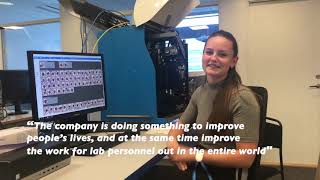
The CellaVision DC-1 is an advanced digital cell morphology analyzer designed to automate and simplify the process of performing blood cell differentials in low-volume hematology laboratories. It leverages high-speed robotics, digital imaging, and artificial intelligence (AI) to automatically locate, capture high-quality images, and pre-classify cells from stained blood smears. The system pre-classifies white blood cells (WBCs) into twelve categories and pre-characterizes red blood cell (RBC) morphology based on six categories, also offering functionality for platelet estimation. This automation significantly reduces manual steps and review time, leading to improved efficiency, quality, and standardization of results compared to traditional microscopy. The CellaVision DC-1 supports both stand-alone and networked installations, enabling remote review and collaboration among medical technologists, supervisors, and morphology experts, which is particularly beneficial for distributed laboratory networks. It is intended for in-vitro diagnostic use in clinical laboratories by trained operators.
Reviewed by Pouyan Golshani, MD — Interventional Radiologist
Key Features
- Automated digital imaging of blood smears
- AI-powered pre-classification of 12 WBC categories and 6 RBC morphologies
- Automated platelet estimation
- Standardized testing process for consistent accuracy
- Reduces manual steps and review time for blood cell differentials
- Supports remote review and collaboration among morphology experts
- Designed specifically for low-volume hematology laboratories
- Bi-directional LIS (Laboratory Information System) support (ASTM)
- Local hard drive storage (up to 1,500 slides) with unlimited secondary storage options
- Built-in quality control features including cell location accuracy test and smear check
Use Cases
- Performing blood cell differentials in low-volume hematology laboratories
- Modernizing and improving blood cell differential processes in small independent labs
- Enabling remote review and expert consultation within distributed laboratory networks
- Standardizing morphological assessments to reduce subjective interpretations
- Improving workflow efficiency and turnaround times for blood smear analysis
- Supporting training and proficiency development in cell morphology (with additional software)
What Physicians Need to Know
The CellaVision DC-1 provides standardized, high-quality digital images and AI-assisted pre-classification of blood cells, which can enhance the accuracy and consistency of differential counts. For challenging cases, the system's remote review capabilities allow for rapid consultation with morphology experts or pathologists, potentially leading to faster diagnoses and treatment decisions. While the AI offers robust support for common cell types, physician oversight remains crucial, especially for rare or complex pathologies like certain leukemias, where the morphologist's final verification is the gold standard.
Seamless integration with existing Laboratory Information Systems (LIS) via bi-directional ASTM communication and Ethernet connectivity ensures efficient data flow and reduces manual entry errors. The ability for multiple CellaVision analyzers to share a database streamlines workflows in distributed laboratory networks, enabling remote review and collaborative diagnostics. This connectivity is key to maximizing the efficiency and quality benefits of digital morphology.
Details
| Category | Lab & Diagnostics, Pathology AI |
| Pricing | Not available publicly — N/A |
| Deployment | On-premise (analyzer with integrated PC), supports networked installation |
| Compliance | |
| BAA Available | Unknown AI-estimated |
| HIPAA Compliant | Unknown AI-estimated |
| FDA Status |
1 AI-estimated The CellaVision DC-1, including the CellaVision DC-1 PPA, received FDA 510(k) clearance (K200595) on October 16, 2020. It is intended for in-vitro diagnostic use in clinical laboratories for differential count of white blood cells (WBC), characterization of red blood cell (RBC) morphology, and platelet estimation. |
| Integrations | |
| EHR | Not specified |
| Specialties | Hematology, Laboratory Medicine, Pathology |
What the Web Says
The CellaVision DC-1 is a digital cell imaging analyzer designed for low-volume and small satellite laboratories, automating and simplifying blood cell differentials. It utilizes AI-based image analysis for pre-classification of cells, which are then reviewed and verified by medical technologists. The system aims to improve efficiency, standardize results, and enable remote collaboration among healthcare professionals.
Overall: MixedStrengths
- Reduces review time for routine films and overall turnaround time (TAT) for smears, especially in distributed laboratory networks.
- Provides high-quality digital imaging and pre-classification of WBCs, RBCs, and platelets using AI, leading to more standardized and consistent results.
- Enables remote review and collaboration with off-site colleagues, supervisors, and pathologists, improving communication and access to morphology experts.
- Compact and easy to operate, making it suitable for smaller labs and as a backup or for specialty testing in larger networks.
- Reduces the physical strain and fatigue associated with traditional manual microscopy.
- Offers educational value through beautiful screenshots and the ability to save cases for sharing and training.
Limitations
- Can struggle with body fluids, especially those with low WBC counts, leading to failed analyses and backlogs.
- Limitations in RBC morphology identification, such as Howell-Jolly bodies, due to the inability to focus up and down on the slide.
- Experienced morphologists may have trouble trusting the technology initially, leading to manual re-review which can negate efficiency gains.
- May make unnecessary workload for cell verification in certain cell classes, particularly those with low positive predictive value (e.g., blasts, promyelocytes, myelocytes, metamyelocytes, plasma cells, nucleated red blood cells).
- Less effective for classifying certain abnormal or rare cell types, such as those found in leukemia samples, where manual review remains the gold standard.
- Some users report issues with inaccurate cell identification, such as eosinophils in the monocyte category or lymphocytes being called blasts, and overcalling certain features like polychromasia or artifacts.
Based on reviews from: Reddit, PubMed, Abacus dx, InPractise, Sysmex America, LabMedica, Tiger Medical, CellaVision (Official Website), ResearchGate, FDA
Last updated: 2026-07-20
Ratings & Reviews
No reviews yet. Be the first to review this tool!
Rate CellaVision DC-1
Press & Coverage
Videos
Product demos, reviews, and walkthroughs for CellaVision DC-1.
CellaVision DC-1
CellaVision