ClariPulmo

by ClariPi  · Based in South Korea → — AI-powered Precision Analytics Solution for Ultra-Low-Dose CT Lung Cancer Screening
Oncology Pulmonology Radiology

Not publicly available
Regulatory Status Disclosed

Overview

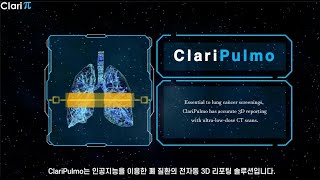
ClariPulmo by ClariPi is a non-invasive, AI-powered image analysis software designed to support the quantification and documentation of pulmonary tissue images from thoracic CT datasets. It is primarily developed for radiologists, pulmonologists, and thoracic imaging specialists working in hospitals, imaging centers, and clinical practices. The software is utilized in clinical settings for lung screenings, particularly for the assessment of emphysema, chronic obstructive pulmonary disease (COPD), and other pulmonary abnormalities.

The tool integrates into existing clinical workflows by offering full compatibility with standard DICOM-compliant CT scanners and Picture Archiving and Communication Systems (PACS). It can be deployed as a standalone desktop application, integrated directly into imaging modalities, or accessed via cloud-based platforms. This allows for automated processing and reporting without disrupting established radiology reading routines.

Notable capabilities of ClariPulmo include:

  • Automated Segmentation: Fully automated segmentation and 3D visualization of the lungs and airways.
  • Lesion Quantification: Precise quantification of low-attenuation areas (LAA) and high-attenuation areas (HAA) using predefined Hounsfield unit thresholds.
  • Advanced Reporting: Generation of color-coded 3D visual representations and structured reports detailing lung volume, LAA/HAA percentages, and lesion locations.
  • Denoising and Normalization: Built-in AI-denoising (via ClariCT.AI) and kernel normalization to ensure consistent, accurate measurements even with ultra-low-dose CT scans or heterogeneous reconstruction conditions.

Reviewed by Pouyan Golshani, MD — Interventional Radiologist

Key Features

  • Automated segmentation of lungs using a pre-trained deep learning model
  • Quantification of low attenuation area (LAA) and high attenuation area (HAA)
  • Provides lung volume, LAA/HAA volume, and percentage in table and pie chart format
  • Offers an overview of the lung in 3D with severity level and percentage
  • Colored overlay of upper, middle, and lower lung zones
  • DICOM compliant for seamless integration with existing CT equipment and PACS
  • Optional denoising function (ClariCT.AI module) for improved quantification of lung CT images
  • Optional Kernel Normalization function for improved quantification of lung CT images
  • Generates easily interpretable reports with color-coded visual representations
  • Lung nodule detection (CE marked only)

Use Cases

  • Support for diagnosis and documentation of pulmonary tissue images (e.g., abnormalities) from CT thoracic datasets
  • Quantitative analysis of lung CT images
  • Automated emphysema reporting
  • Support for reporting on findings related to COVID-19 pneumonia (for documentation, not diagnosis)
  • Facilitating low-dose lung CT screenings
  • Patient condition monitoring and tracking

What Physicians Need to Know

Key Capabilities
Automated segmentation of lungs and quantification of low-attenuation (LAA) and high-attenuation (HAA) areas in CT images. Provides 3D reporting for emphysema and COPD, including lung nodule detection (CE marked only). Features optional denoising (ClariCT.AI module) and kernel normalization for improved image quantification from low-dose CTs. Generates summary reports with color overlays, tables, and charts for analysis results, including lung volume and LAA/HAA percentages.
Clinical Utility
Supports physicians in diagnosing and documenting pulmonary tissue abnormalities from CT thoracic datasets, aiding in emphysema and COPD reporting, and lung cancer screenings. Enhances image clarity and provides quantitative analysis of lesions, designed for reliable detection, quantification, and reporting of lung lesions. Offers patient condition monitoring and tracking, and helps achieve best practices in lung screening by reducing noise-induced bias.
Integration Options
DICOM compliant for seamless integration with existing CT scanners and PACS systems. Vendor-neutral, compatible with any CT scanner brand and model. Can function as a standalone desktop application, be built into modalities, or deployed via cloud services. Integrates with platforms like TeraRecon's Eureka Clinical AI platform and is available on the Nuance AI Marketplace.
Compliance Status
FDA cleared, CE marked, and MFDS (Korea) approved.
Pricing Model
Specific pricing for ClariPulmo is not explicitly detailed, but a 'double solution package at one unit price' is mentioned when combined with ClariCT.AI. For ClariCT.AI, pricing models include one-time license fees and subscriptions based on the number of analyses or installations.
User Experience
User-friendly interface with optional functions for measurement, analysis, and segmentation. Generates easily interpretable reports with color-coded visualizations. Features a fully automated and streamlined workflow with fast processing speeds and simple desktop PC installation.
Support Quality
ClariPi provides contact information (email, phone, fax) for support. The company's foundation in long-term collaborative research between engineering and medical experts suggests a deep understanding of clinical needs and robust product development.
Implementation Complexity
Designed for simple integration with existing DICOM-compliant CT scanners and PACS systems. Its vendor-neutral compatibility eliminates the need for new hardware. Can be implemented as a standalone solution or integrated into existing modalities, with cloud service options available.
Evidence Base
Developed through long-term collaborative research between engineering and medical experts. The underlying ClariCT.AI technology was trained with millions of noise-corrupted CT images. Diagnostic performance shows excellent agreement (PCC: 0.980u20130.983 for HAA, 0.99 for LAA) with expert-established segmentations, including datasets from patients with pneumonia, COVID-19, and healthy individuals. Clinical studies have demonstrated significant radiation dose reduction (up to 1/20th for pediatric patients) with comparable image quality.
Physician Tip

Leverage ClariPulmo for precise, automated quantification of lung lesions (LAA, HAA, nodules) from CT scans, particularly beneficial for lung cancer screening and COPD management. Utilize its denoising and kernel normalization features to improve the diagnostic quality of ultra-low-dose CT images, potentially reducing patient radiation exposure without compromising image clarity. Integrate the 3D reporting and color-coded visualizations into your workflow for enhanced documentation and clearer communication of findings to patients and colleagues. Consider its utility in monitoring and tracking patient conditions over time due to its quantitative analysis capabilities.

ClariPulmo is highly interoperable, designed for seamless integration with existing DICOM-compliant CT scanners and PACS systems, minimizing disruption to current imaging workflows. It can operate as a standalone application, be embedded within modalities, or deployed via cloud services. Strategic partnerships, such as with TeraRecon and availability on the Nuance AI Marketplace, further expand its integration ecosystem.

Details

Category Oncology AI, Radiology & Imaging AI
Pricing Not publicly available
DeploymentStandalone software application; integrates with CT scanners and PACS systems.
Compliance
BAA AvailableUnknown AI-estimated
HIPAA CompliantUnknown AI-estimated
FDA Status 1 AI-estimated

ClariPulmo received U.S. Food and Drug Administration (FDA) 510(k) clearance (K203783) on April 6, 2022. It is classified as radiological image processing software intended to support the quantification of lung CT images.

Integrations
EHR Not specified
Specialties Oncology, Pulmonology, Radiology

What the Web Says

ClariPulmo is a non-invasive AI-powered image analysis software designed to assist physicians in quantifying and documenting lung CT images, particularly for conditions like emphysema and lung nodules. It offers automated segmentation of lungs and airways, and quantification of low-attenuation and high-attenuation areas. The software also includes features like denoising and kernel normalization to improve image quantification, especially in low-dose CT scans.

Overall: Mixed

Strengths

  • AI-powered 3D reporting solution for lung images, including emphysema and lung nodules.
  • Provides accurate and consistent detection and measurement results even with heterogeneous scan conditions due to built-in denoising and kernel normalization.
  • Supports physicians in diagnosis and documentation of pulmonary tissue images.
  • Offers automated segmentation and visualization for lungs and airways, and automatic analysis of low-attenuation and high-attenuation areas.
  • Fully compliant with DICOM standards, allowing seamless integration with CT scanners and PACS systems.
  • Can reduce operating costs as it doesn't require changing existing CT scanners.

Limitations

  • The software is not intended for the diagnosis of pneumonia or COVID-19.
  • No specific reviews from physicians, healthcare IT, tech reviewers, Reddit, G2, or Capterra were found directly for ClariPulmo.
  • General concerns about review platforms like Capterra and G2 regarding authenticity and incentivized reviews exist, which could impact the trustworthiness of any reviews found there.

Based on reviews from: TestDynamics, Claripi, accessdata.fda.gov, G2, Capterra, Quora

Last updated: 2026-07-20

Ratings & Reviews

No reviews yet. Be the first to review this tool!

Rate ClariPulmo

Clinical Value
Ease of Use
Integration
Support & Docs
Value for Money

Press & Coverage

ConcertAI
ConcertAI's TeraRecon Partners with ClariPi to Integrate AI Solutions for Comprehensive Medical Imaging on Eureka Clinical AI Platform
TeraRecon, a ConcertAI company, announced a strategic partnership with ClariPi to integrate ClariPi's AI solutions, including ClariPulmo, into its Eureka Clinical AI platform. This collaboration aims to enhance clinical workflows and improve the identification of lung and breast tissue abnormalities.
2023-07
G-MedTech
TeraRecon Integrates ClariPi's Medical Imaging AI Solutions into its Eureka Clinical AI Platform
This article details the partnership between TeraRecon and ClariPi, highlighting the integration of ClariPi's AI solutions, such as ClariPulmo, into the Eureka platform to improve radiology interpretation efficiency and accuracy. The partnership seeks to bring these AI solutions to a wider range of global customers.
2023-07
PR Newswire
ClariPi to Showcase its AI Medical Imaging Solutions at RSNA 2021
ClariPi announced its participation in RSNA 2021, where it showcased its AI medical imaging solutions, including the introduction of ClariPulmo as a fully automated AI-powered 3D reporting solution for COVID-19 pneumonia and emphysema.
2021-11
ClariPi
ClariPi, '3D Ultra-Low-Dose CT Imaging Technology' Acquired US FDA Approval
ClariPi announced that its product, ClariPulmo, received sales approval from the US Food and Drug Administration (FDA) on April 6, 2022. This approval signifies a significant regulatory milestone for their AI-powered 3D ultra-low-dose CT imaging technology.
2022-04
accessdata.fda.gov
K203783 - Harry Park
This FDA 510(k) Premarket Notification document details the clearance of ClariPulmo (K203783) as a non-invasive image analysis software for quantifying lung CT images. It specifies the software's intended use to support physicians in diagnosing and documenting pulmonary tissue images, excluding the diagnosis of pneumonia or COVID-19.
2022-04
PMC
Impact of Deep Learning 3D CT Super-Resolution on AI-Based Pulmonary Nodule Characterization
This peer-reviewed article discusses the use of ClariPulmo in evaluating pulmonary nodule volumetry and categorization, highlighting how a deep learning-based 3D super-resolution method can improve the accuracy of pulmonary nodule characterization from CT images.
2025-01
Claripi
ClariPulmo | AI-Powered 3D Reporting Solution for LDCT Lung Images
ClariPulmo is presented as an AI-powered 3D reporting solution for LDCT lung images, designed to support physicians in the diagnosis and documentation of pulmonary tissue images through automated segmentation and quantification of lung areas.
unknown
AuntMinnie
Meet the Minnies 2022 semifinal candidates
ClariPulmo AI-powered 3D reporting software by ClariPi was listed as a semifinal candidate in the 'Best New Radiology Device' category for the Minnies 2022 awards.
2022-08

Videos

Product demos, reviews, and walkthroughs for ClariPulmo.

View all on YouTube

Frequently Asked Questions

ClariPulmo is designed to augment, rather than replace, human decision-making in pulmonary care. It can integrate by analyzing existing patient data, such as imaging and pulmonary function tests, to provide diagnostic support or risk stratification. This aims to streamline tasks like documentation and assist with complex diagnoses, allowing physicians to focus more on patient interaction.
As an AI-powered medical device, ClariPulmo would typically require regulatory clearance such as FDA approval in the US or CE Mark in Europe, depending on its risk classification and intended use. The FDA has specific pathways for AI/ML-based Software as a Medical Device (SaMD), which involve rigorous evaluation of safety, effectiveness, and a predetermined change control plan for modifications.
ClariPulmo adheres to strict data privacy protocols, including HIPAA in the US, by implementing robust technical safeguards like encryption and access controls. It requires Business Associate Agreements (BAAs) with third-party vendors and emphasizes the use of de-identified data whenever possible for model training. Regular risk assessments and staff training on secure AI usage are also critical components.
Alternatives to ClariPulmo include traditional diagnostic methods such as manual interpretation of pulmonary function tests and imaging by pulmonologists, which can sometimes have variability in accuracy. Other AI-driven solutions for pulmonary diagnostics also exist, focusing on areas like lung cancer screening, infectious respiratory disease detection, and asthma exacerbation prediction.
Pricing models for healthcare AI solutions like ClariPulmo often include subscription-based fees, consumption-based (pay-per-use), or fixed-cost models, with hybrid options gaining traction. These models aim to balance predictability for budgeting with variable components tied to clinical units or performance. Factors influencing cost include scope, complexity, data quality, and customization.
Like many AI systems, ClariPulmo's algorithms may be susceptible to biases stemming from the training data, potentially leading to disparities in diagnosis or treatment for underrepresented patient populations. Addressing these involves using diverse and representative datasets, ongoing validation in real-world clinical settings, and ensuring transparency in the AI's decision-making process. Human oversight and critical review of AI-generated outputs are essential to mitigate these risks.
Human oversight is strongly recommended, as AI tools like ClariPulmo are intended to augment, not replace, clinical judgment. Physicians remain responsible for verifying AI-generated outputs and making final diagnostic and treatment decisions. Liability for AI-related errors is an evolving area, with current guidelines often placing the burden on clinicians and healthcare organizations to ensure proper validation and use.

Related Tools

Surgical Reality Viewer
Surgical Reality
Oncology AI
Surgical Reality offers CE- and FDA-certified AI-powered 3D medical imaging and surgical planning software that transforms CT scans into patient-specific 3D models for thoracic surgery, enhancing precision in complex lung procedures.
Cancer Copilot
Color Health
Oncology AI
Color Health's Cancer Copilot, built with OpenAI, helps oncologists and clinicians identify and order workup needs for recently diagnosed patients, aiming to speed up time to cancer treatment. This AI tool is designed to expand cancer expertise, speed decisions, and improve care, reducing a two-hour clinical task to 10 minutes with over 95% validated accuracy.
Cercare Medical Oncology Virtual Expert
Cercare Medical
Neurology AI
Cercare Medical's Oncology Virtual Expert is an AI-powered brain tumor segmentation module that provides fast, consistent, and semi-automated tumor analysis from standard MRI data, designed for on-premise deployment within existing clinical radiology workflows.
Synapse Lung Nodule AI
Fujifilm Corporation
Oncology AI
Fujifilm's Synapse Lung Nodule AI is an AI software algorithm for detecting pulmonary nodules on CT imaging, aiming to support early detection and treatment of lung cancer.
RadOncAI
Informai (for RadOncAI product) / Global Network for AI in Radiation Oncology (for RadoncAI.net)
Oncology AI
RadOncAI is an AI solution by Informai designed to optimize and enhance radiation oncology workflows, improving efficiency and accuracy in treatment planning and delivery.
FAITH project (Fatigue Therapy – AI-supported Diagnosis and Therapy of Tumour-associated Fatigue Syndrome)
Fimo Health GmbH (Consortium Leader)
Mental & Behavioral Health AI
The FAITH project is developing an AI-based solution for the diagnosis and therapy of tumor-associated fatigue syndrome in cancer patients. It uses wearable sensors, a smartphone app, and artificial intelligence to provide individualized therapy recommendations and monitor mental health.

See all Oncology AI tools →

More from ClariPi

ClariCT.AI
ClariPi
Radiology & Imaging AI
ClariCT.AI is an FDA-cleared, AI-powered CT image denoising solution by ClariPi, enhancing clarity for low-dose and ultra-low-dose CT scans to improve diagnostic confidence and patient safety.
ClariSIGMAM
ClariPi
Oncology AI
ClariSIGMAM is an AI-powered breast density assessment solution by ClariPi that provides accurate and consistent density estimates from digital mammograms, aiding radiologists in breast tissue composition assessment and breast cancer risk prediction.

View company profile →

Suggest an Edit → | Last Verified: 2026-04-20 | First Added: 2026-04-20

Investors who backed ClariPulmo

Funded through the company that built this tool.

AI Tool Finder
AI-powered search. Results may not be comprehensive.