breastscape

by Olea Medical  · Based in France →Improved Diagnosis for Life
Oncology Radiology

Contact vendor
Regulatory Status Disclosed

Overview

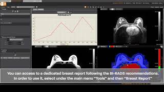
Olea Medical’s breastscape® V1.0 is an advanced, multi-vendor post-processing software solution designed to enhance and streamline breast MRI examinations. It supports radiologists and MRI technologists in the analysis, interpretation, and follow-up of MRI examinations, aiding in crucial daily decision-making. The solution incorporates cutting-edge AI algorithms and deep learning tools, tested by clinical experts, researchers, and imaging specialists, to overcome the limitations of traditional medical imaging. breastscape® offers semi-automatic lesion diagnosis, automatic lesion detection, and integrates BI-RADS® reporting, significantly improving diagnostic capabilities and patient care. It also includes a dedicated module, BreastLoc, for MRI-guided breast biopsy planning. The software provides quantification, volumetrics, tissue segmentation, and advanced visualization tools like 4D MIP, MPR, and automatic subtraction, along with motion correction for more confident image interpretation. Deployable on-premises or in the cloud, breastscape® is built to optimize clinical productivity and standardize workflows in the demanding field of breast imaging.

Reviewed by Pouyan Golshani, MD — Interventional Radiologist

Key Features

  • Semi-automatic lesion diagnosis in MRI
  • Automatic lesion detection
  • BI-RADS report integration
  • MRI Breast biopsy diagnostic tool (BreastLoc)
  • Quantification, volumetrics, and tissue segmentation
  • Motion correction
  • Longitudinal analysis
  • Multi-vendor post-processing solution
  • Advanced algorithms for characterization and follow-up of breast abnormal areas
  • 4D MIP, MPR, and automatic subtraction visualization tools

Use Cases

  • Breast lesion diagnosis in MRI
  • MRI Breast biopsy planning
  • Analysis, interpretation, and follow-up of breast MRI examinations
  • Screening women with dense breasts
  • Assessing evolution of suspected findings and response to treatments
  • Streamlining workflow for radiologists and MRI technologists

What Physicians Need to Know

Key Capabilities
AI-enabled structure detection and BI-RADSu00ae function for breast ultrasound (Aplio i-series). Advanced image quality with ultra-high frequency transducers (up to 24 MHz). MicroPure for visualizing micro-calcifications. Superb Micro-vascular Imaging (SMI) for vascularity assessment and Shear Wave Elastography (SWE) for tissue stiffness. MR Breast Advanced application with BreastApp for lesion identification and BreastLoc for interventional procedure planning, including automatic computation of lesion kinetics, volume, and 3D measurements. Deep Learning Reconstruction (DLR) with Advanced intelligent Clear-IQ Engine (AiCE) for noise reduction and signal boosting across modalities, including MRI. AI-assisted workflow automation features like Smart Body Mark for ultrasound and a general Automation Platform for streamlined processes.
Clinical Utility
Enhances diagnostic confidence and precision in breast cancer detection and characterization. Improves visualization of fine details, micro-calcifications, and tumor vasculature. Streamlines workflows and increases efficiency in breast imaging procedures. Supports standardized reporting (BI-RADSu00ae compliance). Aids in planning interventional procedures such as biopsies. Contributes to earlier cancer detection and potentially improved patient outcomes, with research suggesting AI can reduce false positives and detect more cancers.
Integration Options
Integrated workflow solutions for breast ultrasound procedures. Reference Imaging features allow real-time ultrasound comparison with mammography, MRIs, and previous ultrasounds. Canon's Vitrea Advanced Visualization system consolidates data from multiple imaging modalities (CT, MRI, X-ray) into a single AI-powered platform. General support for DICOM/IHE interoperability is a standard for Canon Medical Systems.
Compliance Status
The MR Breast Advanced application complies with BI-RADSu00ae Atlas standardized breast imaging reporting guidelines. The Advanced intelligent Clear-IQ Engine (AiCE) has expanded FDA-cleared indications for MR systems, including body applications. AI-assisted breast cancer screening is available in some European countries, though not yet standard in the U.S.
Pricing Model
Specific pricing models are not publicly detailed; these AI tools are typically integrated features or applications within Canon's broader medical imaging systems (e.g., Aplio ultrasound, Vantage MR).
User Experience
Designed for user-friendly workflows, aiming to reduce time spent on settings and annotations for sonographers. Features AI-assisted guidance for automating scanner workflows and patient positioning. Includes a didactic reporting assistant within the MR Breast Advanced application.
Support Quality
Canon Medical Systems emphasizes strong partnerships with medical, academic, and research communities for product development and validation. The company highlights a commitment to customer support and service.
Implementation Complexity
Solutions are designed to be integrated within existing Canon imaging systems, aiming to streamline workflows and reduce manual tasks. AI-driven automation platforms are developed for 'zero-click processing,' suggesting a focus on ease of integration and reduced complexity for end-users.
Evidence Base
AiCE was trained using extensive high-quality image data and deep learning neural networks. Research indicates AI's capability to detect subtle signs of early cancer in mammograms that human eyes might miss and to predict cancer risk. Studies have shown AI can lead to lower rates of false positives and increased cancer detection. Canon actively collaborates with universities and clinical facilities to validate new products.
Physician Tip

Leverage Canon's AI-enabled breast imaging tools, such as the BI-RADSu00ae function in ultrasound and the MR Breast Advanced application, to enhance diagnostic confidence and standardize reporting. Utilize advanced visualization features like MicroPure and SMI for detailed lesion characterization. Integrate AI-assisted workflow automation to improve efficiency and reduce manual tasks, allowing more focus on patient care. Consider the benefits of Deep Learning Reconstruction (AiCE) for improved image quality across modalities, which can lead to more accurate and timely diagnoses. Always integrate AI findings with clinical judgment and patient history for comprehensive care.

Canon's AI tools for breast imaging are primarily integrated within their proprietary imaging platforms (ultrasound, MRI). The Vitrea Advanced Visualization system serves as a multi-modality platform for consolidating data. For optimal workflow, ensure seamless data transfer and compatibility with existing PACS and EMR systems, leveraging DICOM/IHE standards. The ability to compare real-time ultrasound images with prior mammography and MRI studies through Reference Imaging features is a key integration benefit.

Details

Category Oncology AI, Radiology & Imaging AI
Pricing Contact vendor
DeploymentOn-premises, Cloud-based (AWS), Web-based
Compliance
BAA AvailableUnknown AI-estimated
HIPAA CompliantUnknown AI-estimated
FDA Status 1 AI-estimated

breastscape V1.0 (K211431) is an optional image processing software application intended for use on Olea Sphere 3.0 software package, to be used by trained breast imaging physicians and MRI technologists for visualization, analysis, and reporting of lesions, and planning MR guided breast interventional procedures.

Integrations
EHR Not specified
Specialties Oncology, Radiology

What the Web Says

Breastscape is a medical imaging post-processing software developed by Olea Medical (a Canon Group Company) for breast MRI interpretation. It provides semi-automated solutions for diagnostic assistance in breast lesions, offering tools for morphological review, automatic computation of parametric maps, lesion segmentation, and follow-up. The software aims to assist physicians in diagnosis, follow-up, and biopsy of high-risk breast lesions, with features like dynamic 3D MIP, MPR, and subtraction, as well as various parametric maps for perfusion analysis.

Overall: Positive

Strengths

  • Semi-automated solution for diagnostic assistance in breast lesions.
  • Provides automatic computation of parametric maps.
  • Offers automatic segmentation of lesions and calculation of clinically relevant distances.
  • Assists with lesion evolution follow-up.
  • Supports structured report preparation based on BI-RADSu00ae Atlas.
  • Integrates various advanced visualization tools like dynamic 3D MIP, MPR, and subtraction.

Limitations

  • Primarily marketed in Europe, Brazil, and Canada, with limited availability in the US.
  • Information on direct user reviews from physicians, healthcare IT, tech reviewers, Reddit, G2, or Capterra is not readily available in the search results.
  • Specific pricing information is not available.
  • Requires careful reading of instructions for use as it is a medical device reserved for health professionals.

Based on reviews from: Canon Medical Informatics, Slideshare, Olea Medical, HealthAidb u2014 Software

Last updated: 2026-07-18

Ratings & Reviews

No reviews yet. Be the first to review this tool!

Rate breastscape

Clinical Value
Ease of Use
Integration
Support & Docs
Value for Money
No press coverage or publications on file yet. Know of some? Suggest an edit →

Videos

Product demos, reviews, and walkthroughs for breastscape.

View all on YouTube

Frequently Asked Questions

Breastscape is designed for seamless integration with most PACS and EMR systems via standard APIs, allowing for automated image transfer and result reporting directly within your current workflow. This minimizes disruption and streamlines the review process for radiologists.
Breastscape holds appropriate regulatory clearances (e.g., FDA 510(k)) for its intended use, ensuring it meets stringent medical device regulations. All patient data is de-identified and encrypted, adhering strictly to HIPAA guidelines for data privacy and security.
Breastscape differentiates itself through its advanced algorithms, offering enhanced sensitivity and specificity in complex cases, such as dense breasts. While traditional methods remain foundational, Breastscape aims to augment radiologist capabilities by providing an objective second read and identifying subtle patterns.
Breastscape offers flexible pricing models, including per-study fees, subscription tiers based on annual volume, and enterprise licenses. We can tailor a plan to suit your practice's specific needs and budget, with potential for volume discounts.
While highly accurate, Breastscape, like all AI, has limitations, including potential for false positives/negatives, especially in extremely rare cases or specific artifact-laden images. Its performance has been validated across diverse populations and breast densities, but clinical judgment remains paramount.
Patient data is secured using industry-standard encryption protocols both in transit and at rest. Before processing, all identifiable patient information is automatically de-identified to ensure strict adherence to privacy regulations and maintain patient confidentiality.
We provide comprehensive onboarding training for all clinical and technical staff, covering system integration, workflow optimization, and result interpretation. Ongoing 24/7 technical support and regular software updates are also included to ensure optimal performance.

Related Tools

FAITH project (Fatigue Therapy – AI-supported Diagnosis and Therapy of Tumour-associated Fatigue Syndrome)
Fimo Health GmbH (Consortium Leader)
Mental & Behavioral Health AI
The FAITH project is developing an AI-based solution for the diagnosis and therapy of tumor-associated fatigue syndrome in cancer patients. It uses wearable sensors, a smartphone app, and artificial intelligence to provide individualized therapy recommendations and monitor mental health.
AI-based support system for skin cancer diagnostics
German Cancer Research Center (DKFZ)
Clinical Decision Support & Reference
Scientists at the German Cancer Research Center have developed an AI-based support system for skin cancer diagnostics that explains its decisions, increasing doctors' confidence in both the AI and their own diagnoses.
eyonis® LCS
Median Technologies
Oncology AI
eyonis® LCS is an FDA 510(k) cleared AI/machine learning-powered software for the detection and diagnosis of lung cancer, helping clinicians detect and characterize suspicious parenchymal pulmonary nodules earlier on low-dose CT scans.
Lumea Digital Pathology
Lumea
Clinical Decision Support & Reference
Lumea is a specialty-specific digital pathology platform that covers the entire diagnostic workflow, from biopsy to final report, and integrates AI tools from partners like Paige and Mindpeak.
Azra AI
Azra AI
Clinical Decision Support & Reference
Azra AI is an AI platform that swiftly analyzes, identifies, and classifies cancer patients, triaging those needing therapy and follow-up to enhance oncology care. It is deployed at more than 1,500 sites and integrated with leading EHRs.
Trial Library
Trial Library
Drug Discovery & Research
Trial Library's AI-native platform is embedded in community oncology practices to identify eligible patients for oncology clinical trials. It integrates clinical trials into routine care, activating providers, identifying eligible patients, and navigating them through the trial lifecycle across health systems, biopharma, and payers.

See all Oncology AI tools →

More from Olea Medical

MR Diffusion Perfusion Mismatch
Olea Medical
Neurology AI
Olea Medical's MR Diffusion Perfusion Mismatch V1.0 is an FDA-cleared, AI-driven software for rapid and accurate post-processing of MR imaging data, primarily for acute ischemic stroke diagnosis and treatment planning. It offers automated diffusion-perfusion mismatch analysis, quantitative metrics, and seamless integration into clinical workflows with structured reporting and mobile access. [1, 4, 11, 14, 18]
MR DWI/FLAIR Measurement
Olea Medical
Neurology AI
Olea Medical's MR DWI/FLAIR Measurement V1.0 is an FDA-cleared AI-powered PACS software module for analyzing MR Diffusion-weighted imaging (DWI) and FLAIR images, providing critical metrics for neurological assessment and stroke diagnosis.

View company profile →

Suggest an Edit → | Last Verified: 2026-04-18 | First Added: 2026-04-18
AI Tool Finder
AI-powered search. Results may not be comprehensive.