EFAI ChestSuite XR Pneumothorax Assessment System
Overview
The EFAI ChestSuite XR Pneumothorax Assessment System is a radiological computer-assisted triage and notification software system developed by Ever Fortune AI Co.. It utilizes deep learning techniques to automatically analyze Posteroanterior (PA) chest X-rays, identifying suspected findings of pneumothorax. The system is designed to send notification messages to Picture Archiving and Communication Systems (PACS) or workstations, facilitating worklist prioritization and triage for medical professionals.
This software workflow tool is intended to aid the clinical assessment of adult (22 years of age or older) PA Chest X-Ray cases that exhibit features suggestive of pneumothorax. It provides a passive notification to radiologists, indicating cases that may benefit from prioritization, but it does not mark, highlight, or direct users’ attention to specific locations on the original chest X-ray. The system is not intended for stand-alone clinical decision-making, nor is it meant to rule out pneumothorax or otherwise preclude a comprehensive clinical assessment of X-ray cases. The EFAI ChestSuite XR Pneumothorax Assessment System demonstrates high performance with a reported sensitivity of 97% and specificity of 98% for pneumothorax detection. Deployment is recommended within a local network with an existing hospital-grade IT system, requiring installation on a specialized server that supports deep learning processing.
Reviewed by Pouyan Golshani, MD — Interventional Radiologist
Key Features
- Radiological computer-assisted triage and notification software
- Utilizes deep learning techniques for automated analysis of PA chest x-rays
- Identifies suspected findings of pneumothorax
- Sends notification messages to PACS/workstation for worklist prioritization or triage
- Aids clinical assessment of adult (22+ years) Posteroanterior (PA) view Chest X-Ray cases
- Not intended for stand-alone clinical decision-making
- Not intended to direct attention to specific portions or anomalies of an image
- Not intended to rule out pneumothorax or otherwise preclude clinical assessment
- High sensitivity (97%) and specificity (98%) for pneumothorax detection
- Recommended deployment in a local network with an existing hospital-grade IT system
Use Cases
- Prioritization and triage of adult PA chest X-ray images for suspected pneumothorax
- Aid for radiologists and clinicians in identifying potential pneumothorax cases
- Improving workflow efficiency in radiology departments by flagging critical cases
What Physicians Need to Know
This AI tool is designed to assist in the triage and prioritization of chest X-rays for suspected pneumothorax, not to serve as a standalone diagnostic solution. Radiologists should use the passive notifications to prioritize their worklist and perform a comprehensive review of all images. The system is specifically validated for adult PA and AP chest X-rays. It does not provide image-level markings or highlights, requiring the radiologist to confirm the presence and location of pneumothorax. Always integrate AI findings with full clinical context and patient history.
The EFAI ChestSuite XR Pneumothorax Assessment System is designed for integration within a local network with existing hospital-grade IT systems. It requires installation on a specialized server capable of deep learning processing. Seamless integration with PACS/workstations is achieved via DICOM standards for image input and notification outputs, facilitating worklist prioritization. Configuration of input and output destinations is typically managed by the manufacturer.
Details
| Category | Radiology & Imaging AI, Triage & ER/ICU AI |
| Pricing | Contact for pricing |
| Deployment | On-premise (local network, specialized server) |
| Compliance | |
| BAA Available | Unknown AI-estimated |
| HIPAA Compliant | Unknown AI-estimated |
| FDA Status |
1 AI-estimated Cleared by the U.S. FDA on May 31, 2022, under 510(k) K221552. It is classified as a Radiological Computer Aided Triage and Notification Software, intended to aid in the clinical assessment of adult (22 years of age or older) Posteroanterior (PA) view Chest X-Ray cases with features suggestive of pneumothorax. |
| Integrations | |
| EHR | Not specified |
| Specialties | Critical Care, Emergency Medicine, Radiology |
What the Web Says
The EFAI ChestSuite XR Pneumothorax Assessment System is an AI-powered software tool designed to help clinicians prioritize chest X-ray cases suspected of pneumothorax. It uses deep learning to analyze posteroanterior chest X-rays and provides case-level notifications within existing PACS/workstation workflows, aiming for faster identification and assessment of patients. The system is intended to aid in triage and clinical decision-making, but not for stand-alone diagnosis.
Overall: PositiveStrengths
- High sensitivity (0.97) and specificity (0.98) for pneumothorax detection.
- Fast performance, with an average analysis time of approximately 23 seconds per case.
- Integrates with existing PACS/workstation workflows, providing case-level notifications for prioritization.
- Aids in triage and supports clinical decision-making, potentially reducing delays in diagnosis.
- Consistent and reliable performance across various X-ray manufacturers and confounding conditions.
- Can improve diagnostic accuracy, especially for less experienced clinicians, and narrow the performance gap between junior and senior readers.
Limitations
- Not intended for stand-alone diagnosis; requires human clinician review.
- Does not mark specific image areas or direct attention to particular anomalies.
- Performance may decrease with multiple findings or for smaller targets.
- Can produce false-positive results, which could lead to unnecessary imaging and costs.
- General-purpose AI models, while showing promise, may have distinct diagnostic biases and varying performance profiles.
- Some AI models for pneumothorax detection have shown reduced performance on real-world clinical images compared to curated datasets.
Based on reviews from: FDA Radiology AI Device (EFAI ChestSuite XR Pneumothorax Assessment System), Ever Fortune AI | Radiology AI Companies - X-ray Interpreter, accessdata.fda.gov (Ever Fortune AI Co., Ltd. 510(k) Premarket Notification), accessdata.fda.gov (Ever Fortune.AI, Co., Ltd. 510(k) Premarket Notification), Muriel Steele Society (Artificial Intelligence Tool for Reads Chest X-Rays Approved by FDA), EverFortune.AI (ChestSuite), Semantic Scholar (Multidisciplinary Evaluation of an AI-Based Pneumothorax Detection Model), GE Healthcare (Critical Care Suite on-device AI), PMC (Evaluating AI Models for Pneumothorax Detection on Chest Radiographs: Diagnostic Accuracy and Clinical Trade-Offs), PMC (Impact of AI Assistance in Pneumothorax Detection on Chest Radiographs Among Readers of Varying Experience), medRxiv (Evaluation of an artificial intelligence model for detection of pneumothorax and tension pneumothorax on chest radiograph), Diagnostic Imaging (FDA-Cleared AI Triage Software for Chest X-Rays Offers Enhanced Detection of Pleural Effusion and Pneumothorax), FDA Clears Critical Care Suite With AI-Powered Pneumothorax Detection, Radiologists Outperformed AI in Identifying Lung Diseases on Chest X-ray, PubMed (Comparison of emergency physicians and artificial intelligence models in pneumothorax detection: A multi-reader retrospective study), Intel (GE Healthcare Accelerates Pneumothorax Detection by Embedding AI with the X-ray System), PLOS Medicine (Automated detection of moderate and large pneumothorax on frontal chest X-rays using deep convolutional neural networks: A retrospective study), CTV News (AI system more accurately identifies collapsed lungs using chest x-rays), Artificial Intelligence Tool for Reads Chest X-Rays Approved by FDA
Last updated: 2026-07-18
Ratings & Reviews
No reviews yet. Be the first to review this tool!
Rate EFAI ChestSuite XR Pneumothorax Assessment System
Videos
Product demos, reviews, and walkthroughs for EFAI ChestSuite XR Pneumothorax Assessment System.
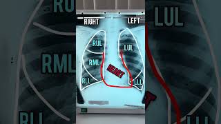
Pneumothorax on a Chest X-Ray #medicine