Synapse 3D Base Tools

by FUJIFILM Corporation  · Based in Japan → — Advanced Visualization and Analysis for Medical Imaging
Neurosurgery Orthopedics Radiology

Contact for pricing
Regulatory Status Disclosed

Overview

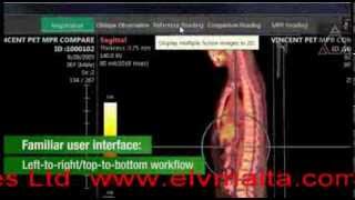
Synapse 3D Base Tools (V7.0) is an advanced medical imaging software from FUJIFILM designed to provide comprehensive tools for advanced image visualization and analysis. It assists trained medical professionals in reading, interpreting, reporting, and planning treatment using images from various modalities such as CT, MRI, X-ray, ultrasound, and nuclear medicine. The software offers a state-of-the-art system that supports not only Radiological and Cardiological Departments but also aids in clinical and surgical workflows. Leveraging Fujifilm’s proprietary Image Intelligence™ technology, Synapse 3D ensures fast, reliable, and accurate processing. It utilizes thin client technology, allowing for enterprise-wide operability and remote client access via VPN or SSL configurations, reducing the need for proprietary hardware. The platform is designed to optimize workflows, enhance diagnostic power, and support interdisciplinary collaboration by providing a unified user interface and shared tools across its modules.

Reviewed by Pouyan Golshani, MD — Interventional Radiologist

Key Features

  • 2D/3D/4D Viewer
  • Multimodality Image Fusion
  • AI-powered Segmentation (Deep Learning)
  • Multi-Planar Reconstructions (MPR)
  • Volume Rendering
  • Thin Client Technology / Web-based Access
  • DICOM Compliant
  • Image Intelligenceu2122 (Proprietary AI Technology)
  • Snapshot Workflows for Collaboration
  • Server-based Image Rendering

Use Cases

  • Diagnostic interpretation and reporting
  • Treatment planning
  • Preoperative simulation
  • Advanced image analysis for various body parts and modalities (CT, MR, CR, US, NM, PT, XA)
  • Interdisciplinary collaboration and workflow optimization
  • Oncology (tumor analysis, RECIST & PERCIST tracking)

What Physicians Need to Know

DICOM Support & Standards
Synapse 3D Base Tools is DICOM compliant, accepting medical images from various modalities (CT, MR, CR, US, NM, PT, XA) and communicating with PACS systems via DICOM standard for data retrieval and storage. It supports DICOM TCP/IP Network Communication.
PACS Integration Method
The tool integrates directly into Synapse PACS Viewer, enabling a 'zero-click' workflow with automatic study processing. It can also integrate with any PACS via desktop integration, facilitating DICOM transfer of images, data, and reports. This integration eliminates the need for separate workstations or logins.
Reading Room Workflow Impact
Synapse 3D is designed to enhance operational efficiency, optimize interdisciplinary collaboration, and streamline workflows. It offers 'zero-click' preprocessing, automated prerendering, and snapshot technology for seamless work hand-off. Its unified user interface and thin-client technology support remote access and reduce the need for multiple workstations.
AI Model Architecture (deep learning approach)
Synapse 3D is powered by Fujifilm's proprietary 'Image Intelligence' technology, utilizing award-winning algorithms and image recognition modules. Recent versions incorporate 'AI-Enabled Workflows' and 'AI assistance' for tasks like anatomical region identification and pathological significance. Mentions of 'Convolutional Network' and 'AI-Driven Detection Systems' (e.g., Synapse AI Orchestrator) suggest the integration of deep learning approaches for complex computations and workflow efficiency.
FDA Clearance Pathway (510k/De Novo)
Synapse 3D Base Tools has received 510(k) clearance from the FDA. Recent clearances include K221677 (V6.6 on 11/10/2022) and K203103 (V6.1 on February 9, 2021). It is classified as a Class II medical device under regulation 21 CFR 892.2050.
Supported Modalities (CT/MRI/X-ray/US)
The tool accepts DICOM compliant medical images from a wide range of modalities including CT, MR, CR, US (Ultrasound), NM (Nuclear Medicine), PT (PET), and XA (Angiography). It is not intended for primary diagnostic interpretation of mammography images.
Sensitivity & Specificity Data
Specific sensitivity and specificity data are not explicitly provided for Synapse 3D Base Tools, as it functions primarily as an advanced visualization and post-processing tool for clinical decision support, rather than a standalone diagnostic AI tool that generates a definitive positive/negative result.
RSNA/ACR Validation
Fujifilm has consistently showcased Synapse 3D at RSNA annual meetings, highlighting its advanced visualization, analysis tools, and integration capabilities. There is no explicit mention of formal ACR validation in the provided information.
Physician Tip

Leverage Synapse 3D's 'zero-click' workflow and snapshot technology for efficient image processing and seamless collaboration across specialties. Utilize its comprehensive 3D/4D visualization and advanced analysis tools for enhanced diagnostic confidence, surgical planning, and patient education. The thin-client architecture allows for flexible access from various locations, improving accessibility and interdisciplinary review. While not a primary diagnostic AI, its AI-enabled workflows can assist in identifying anatomical regions and streamlining tasks. Remember that it is not intended for primary mammography interpretation.

Synapse 3D offers robust integration with Fujifilm's Synapse PACS and Synapse Cardiovascular, providing a unified platform and eliminating the need for separate workstations. It also integrates with any PACS via DICOM desktop integration, allowing for flexible deployment within existing IT infrastructures. The web-based thin-client application reduces bandwidth usage and supports enterprise-wide operability, including remote access via VPN or SSL configurations. Its ability to consolidate data from disparate information systems into a single database enhances efficiency.

Details

Category Radiology & Imaging AI, Surgical AI
Pricing Contact for pricing — Custom pricing options available; contact vendor for details.
DeploymentWindows server/client configuration; Thin client technology; Virtualized environment (VMware ESX servers); Cloud-enabled design.
Compliance
BAA AvailableUnknown AI-estimated
HIPAA CompliantUnknown AI-estimated
FDA Status 1 AI-estimated

Synapse 3D Base Tools (V7.0) is advanced medical imaging software from FUJIFILM designed to assist trained medical professionals in reading, interpreting, reporting, and treatment planning for DICOM compliant images from various imaging devices including CT, MR, CR, US, NM, PT, and XA. It provides powerful 3D viewing tools, image fusion, and segmentation powered by deep learning, enhancing diagnostic workflows and treatment planning for adult patients.

Integrations
EHR Not specified
Specialties Neurosurgery, Orthopedics, Radiology

What the Web Says

Fujifilm Synapse 3D Base Tools is an advanced visualization and analysis software for medical imaging, offering a comprehensive suite of tools for radiologists, cardiologists, surgeons, and other clinical professionals. It aims to enhance diagnostic confidence, aid in treatment planning, and improve workflow efficiency through its integrated platform and specialized applications.

Overall: Mixed

Strengths

  • Comprehensive suite of over 50 clinical applications for various specialties, including cardiology, pulmonology, neurology, gastroenterology, oncology, and urology.
  • Seamless integration with Synapse PACS and Synapse Cardiovascular, allowing for a unified workflow and eliminating the need for separate workstations or logins.
  • Thin-client technology and web-based access, reducing capital investment in high-specification terminals and enabling remote access.
  • Utilizes Fujifilm's proprietary Image Intelligenceu2122 algorithms for fast, reliable, and accurate image recognition and processing.
  • Features like snapshot workflows and application linking streamline the process between pre-processing, reading images, and surgical planning, improving efficiency and interdisciplinary collaboration.
  • Aids in surgical planning and patient education through 3D modeling and visualization, potentially leading to improved patient safety and surgical effectiveness.

Limitations

  • Some users have reported the Synapse system (including PACS) can be glitchy at times, leading to unexpected logouts.
  • Routine slowness issues have been reported, making the system unusable for large studies.
  • The user experience (UX) design may be considered a bit behind the curve, though users can adapt to it over time.
  • Older versions of Synapse clients (e.g., Synapse 4) are not supported on newer operating systems like Windows 11, leading to functionality issues.
  • Some Reddit users have expressed difficulty with free-rotating X-ray images, with the system often limiting rotation to 90-degree increments.
  • Compared to alternatives, Synapse 3D is sometimes rated lower in terms of reliability and ease of use by G2 reviewers, with alternatives like 3D Slicer, Simpleware, and Invivo5 receiving higher ratings.

Based on reviews from: Fujifilm Healthcare Solutions, Imaging Technology News, G2, Reddit, Fujifilm (United Kingdom), Medical Outfitters, Fujifilm, Kebomed, PR Newswire, EYM Solutions, PubMed, Capterra

Last updated: 2026-07-18

Ratings & Reviews

No reviews yet. Be the first to review this tool!

Rate Synapse 3D Base Tools

Clinical Value
Ease of Use
Integration
Support & Docs
Value for Money

Press & Coverage

X-ray Interpreter
Synapse 3D Base Tools (V7.2) | FDA Radiology AI Device
FUJIFILM's Synapse 3D Base Tools (V7.2) is advanced medical imaging software designed to assist trained professionals in reading, interpreting, reporting, and planning treatment using images from various modalities. It provides powerful 3D viewing tools, image fusion, and segmentation powered by deep learning, enhancing diagnostic workflows and treatment planning for adult patients.
2026-04
Innolitics
Q2 2026 AI/ML FDA Clearances and De Novos - Innolitics
This report on Q2 2026 AI/ML FDA clearances mentions Fujifilm's Synapse 3D Base Tools (V7.2) as one of the authorized devices, highlighting its role in radiology. The article discusses trends in AI/ML device authorizations and review timelines.
2026-07
X-ray Interpreter
Synapse 3D Base Tools (V7.0) | FDA Radiology AI Device - X-ray Interpreter
FUJIFILM's Synapse 3D Base Tools (V7.0) is advanced medical imaging software that assists professionals in interpreting medical images from various modalities, offering 3D viewing, image fusion, and deep learning-powered segmentation. This version received FDA approval in May 2025.
2025-05
Future Market Insights
Radiology AI Market Report 2025-2030, By Offering, Function, and Geo
This market report highlights Fujifilm's Synapse 3D Base Tools as a key innovation in the radiology AI market, enabling automated fusion of CT, MR, and PET data into synchronized 3D renderings for volumetric tumor tracking and dual energy analysis.
2025-11
accessdata.fda.gov
K243762 - 510(k) Premarket Notification - FDA
This FDA 510(k) premarket notification details the clearance of Synapse 3D Base Tools (V7.0) by Fujifilm Corporation, outlining its intended use as medical imaging software for trained professionals to aid in reading, interpreting, reporting, and treatment planning.
2025-05
accessdata.fda.gov
K254189 - 510(k) Premarket Notification - FDA
This FDA 510(k) premarket notification pertains to Synapse 3D Base Tools (V7.2), an updated software version intended for medical imaging interpretation and treatment planning, accepting DICOM compliant images from various devices.
2026-04
accessdata.fda.gov
K203103 - 510(k) Premarket Notification - FDA
This FDA 510(k) premarket notification describes Synapse 3D Base Tools (V6.1) as medical imaging software for trained professionals to aid in reading, interpreting, reporting, and treatment planning using DICOM compliant images.
2021-02
X-ray Interpreter
ECHELON Synergy - Physician Ai Tools - Medical AI Tools for ...
FUJIFILM Synapse 3D Base Tools is listed as advanced medical imaging software within the context of other Fujifilm AI devices, emphasizing its role in radiology and imaging AI.
2026-04

Videos

Product demos, reviews, and walkthroughs for Synapse 3D Base Tools.

View all on YouTube

Frequently Asked Questions

Synapse 3D incorporates AI-driven detection systems to accurately identify anatomical regions and areas of pathological significance, thereby enhancing diagnostic precision and confidence. It provides a comprehensive suite of advanced visualization and image analysis tools, including 3D rendering, vessel analysis, and organ-focused visualization, across various clinical specialties. This technology aims to streamline routine tasks and assist with image interpretation, allowing physicians to dedicate more focus to patient care.
Fujifilm Synapse 3D is engineered with robust data security protocols and adheres to stringent HIPAA standards to protect sensitive patient information. It ensures the confidentiality, integrity, and availability of patient data, with some Synapse products also holding DIACAP-certification from the U.S. Department of Defense. The system securely retrieves and stores image data through DICOM standard connections to medical devices and PACS.
Synapse 3D is designed for seamless integration with Fujifilm's Synapse PACS and Synapse Cardiovascular systems, offering 'zero-click' access to advanced visualization tools directly within the workflow. It also supports integration with other third-party PACS through desktop integration, enabling the transfer of images, data, and reports via DICOM. This enterprise-wide operability facilitates efficient interdisciplinary collaboration and allows access from various connected devices.
While Synapse 3D offers advanced visualization and AI-enhanced workflows, it is intended to assist trained medical professionals in interpretation, reporting, and treatment planning, and is not for the primary diagnostic interpretation of mammography images. Its overall performance can also be influenced by the seamless integration with upstream systems like PACS and VNA, as inconsistencies in data routing or access may affect the user experience. The accuracy of its AI-driven detection systems is continuously validated through ongoing performance testing.
Several alternatives are available for medical 3D visualization and advanced imaging, including OsiriX, which offers 2D, 3D, and 4D visualization with advanced post-processing tools. Other options include D2P for 3D model segmentation and printing, PICOM365 as a cloud-based platform, and enterprise imaging solutions like Syngo Carbon from Siemens Healthineers. Additionally, 3D Slicer, Simpleware, and Materialise Mimics Care Suite provide various features for medical image processing and surgical planning.
The pricing and licensing details for Synapse 3D Base Tools are generally not publicly disclosed and are often structured within broader enterprise agreements. This lack of public transparency can make initial budget comparisons challenging, often necessitating direct consultation with Fujifilm to determine specific module requirements and associated costs.

Related Tools

Surgical Reality Viewer
Surgical Reality
Oncology AI
Surgical Reality offers CE- and FDA-certified AI-powered 3D medical imaging and surgical planning software that transforms CT scans into patient-specific 3D models for thoracic surgery, enhancing precision in complex lung procedures.
Oxipit ChestLink
Oxipit (part of Sectra group)
Clinical Decision Support & Reference
Oxipit ChestLink is the world's first CE Class IIb-certified autonomous AI application for healthy chest X-ray reporting, capable of identifying normal studies with 99.9% sensitivity. It automates the reporting of normal chest X-rays, reducing radiologist workload and improving efficiency.
Cercare Medical Oncology Virtual Expert
Cercare Medical
Neurology AI
Cercare Medical's Oncology Virtual Expert is an AI-powered brain tumor segmentation module that provides fast, consistent, and semi-automated tumor analysis from standard MRI data, designed for on-premise deployment within existing clinical radiology workflows.
CARPL.ai
CARPL.ai
Developer Tools & APIs
CARPL.ai is a vendor-neutral Artificial Intelligence (AI) platform that allows radiologists to access, assess, and integrate radiology AI solutions in their clinical practice. It provides a single user interface, a single data channel, and a single procurement channel for the testing, deployment, and monitoring of AI solutions in clinical radiology workflows.
CoLumbo C-Spine
Smart Soft Healthcare AD
Clinical Decision Support & Reference
CoLumbo C-Spine is an AI-powered software that processes and analyzes cervical spine MRI exams, assisting clinicians in diagnosis and assessment.
SubtleHD-CT
Subtle Medical, Inc.
Radiology & Imaging AI
Subtle Medical's SubtleHD-CT is an AI software designed to enhance the quality and resolution of CT scans, reducing noise and improving image clarity. [7]

See all Radiology & Imaging AI tools →

More from Fujifilm

ECHELON Synergy
Fujifilm Corporation
Radiology & Imaging AI
ECHELON Synergy is Fujifilm's AI-powered 1.5T MRI system, cleared by the FDA, that utilizes Deep Learning Reconstruction (DLR) to deliver high-quality images, faster scan times, and enhanced patient comfort through features like a wide bore and motion artifact reduction technologies. [2, 7, 18, 22]
EW10-EC02 Endoscopy Support Program
FUJIFILM Corporation
Gastroenterology AI
The FUJIFILM EW10-EC02 Endoscopy Support Program, part of the CAD EYE system, is an AI-powered software that provides real-time visual and auditory alerts to aid endoscopists in detecting colonic polyps and adenomas during colonoscopy procedures.
SCENARIA View Phase
FUJIFILM Corporation
Cardiology AI
FUJIFILM SCENARIA View Phase is an AI-powered CT system offering advanced imaging, significant dose reduction, and streamlined workflows for comprehensive diagnostic radiology, including cardiac and low-dose lung cancer screening.
Synapse Lung Nodule AI
Fujifilm Corporation
Oncology AI
Fujifilm's Synapse Lung Nodule AI is an AI software algorithm for detecting pulmonary nodules on CT imaging, aiming to support early detection and treatment of lung cancer.

View company profile →

Suggest an Edit → | Last Verified: 2026-04-18 | First Added: 2026-04-18
AI Tool Finder
AI-powered search. Results may not be comprehensive.