MR Diffusion Perfusion Mismatch

by Olea Medical  · Based in France → — Improve Diagnosis for Life
Interventional Radiology Neurology Radiology

Regulatory Status Disclosed

Overview

Olea Medical’s MR Diffusion Perfusion Mismatch V1.0 is an intelligent, automated software solution designed for rapid and accurate post-processing of Magnetic Resonance (MR) imaging data, particularly crucial in acute ischemic stroke diagnosis. [1, 11, 14, 18, 22] This AI-driven tool seamlessly integrates into clinical workflows, providing instant qualitative and quantitative parameters to enhance collaboration and optimize evaluation methodology for various pathologies. [1, 4, 5, 18]

The software offers automated diffusion-perfusion mismatch analysis, which is vital for identifying the infarct core and penumbra in stroke patients, aiding in critical treatment decisions. [11, 14, 15, 18, 22, 25] It provides quantitative analysis of hypoperfused tissue volume, ADC lesion volume, and DWI-FLAIR volumes mismatch. [1, 14, 18, 22] Built on proprietary Bayesian algorithms and advanced optimization methods, Olea Medical’s solutions are recognized for their speed, stability, and efficacy, transforming MR and CT datasets into clear, actionable insights within a unified workflow. [4, 5, 8, 17, 18]

Key functionalities include no-click access to quantitative information, CBV corrected and permeability maps computation, ADC mapping, and structured reporting with automatic export to PACS and email. [1, 4, 18] The system is vendor-neutral, compatible with a wide range of MR and CT scanners, and supports an enhanced intuitive workflow. [1, 5, 6, 19, 20] A dedicated mobile application further supports neuroradiologists by delivering real-time alerts, notifications, and patient image overviews, enabling faster and more consistent decisions in time-sensitive emergency settings. [4, 18]

Reviewed by Pouyan Golshani, MD — Interventional Radiologist

Key Features

  • Automated Diffusion-Perfusion Mismatch analysis
  • Quantitative analysis of stroke parameters (e.g., hypoperfused tissue volume, ADC lesion volume)
  • Fast and accurate image processing
  • Seamless integration into clinical workflows (PACS, email)
  • Vendor-neutral compatibility with MR and CT scanners
  • AI-driven detection, quantification, and structured reporting
  • Dedicated mobile application for real-time alerts and image overview
  • Proprietary Bayesian algorithms for improved signal processing
  • DWI-FLAIR volumes mismatch assessment for infarct dating
  • Automated restrictive diffusion and threshold-based hypo-perfused volume extraction

Use Cases

  • Acute ischemic stroke diagnosis and treatment decision support
  • Neurovascular emergency solutions for rapid triage
  • Brain tumor diagnosis and assessment (as part of broader Olea solutions)
  • Optimizing clinical productivity and radiology workflow
  • Longitudinal analysis and response-to-treatment assessment
  • Functional brain connectivity assessments (related to broader neuro imaging capabilities)

What Physicians Need to Know

Stroke Detection Speed
Olea Medical's products can automatically generate stroke maps in a matter of seconds, providing a reliable and quick tool for detecting stroke. The autoNeurovascular solution is designed to optimize the hospital workflow for patients suspected of stroke, allowing clinicians to use the first available imaging method (CT or MRI) without losing time. AI-based systems can significantly reduce the time to diagnosis and treatment, which is critical for improving patient outcomes.
Brain MRI/CT Analysis
The tool offers comprehensive neuroimaging analysis for both CT and MRI workflows, assisting in decision-making for pre- and post-treatment environments. It provides rapid brain imaging information for differentiating between benign and malignant conditions, and for a clearer view of therapeutic response and disease progression. The software allows for sequence fusion, enhanced contrast, subtle anomaly detection, and functional mapping reconstruction, which helps identify the ischemic penumbra and distinguish it from the necrotic core. It also offers automated lesion segmentation, volume calculation, and perfusion mapping to speed up decision-making. The Neuro INSIGHT solution streamlines radiologists' workflow for neurological disorders by providing detailed qualitative and quantitative parameters for characterizing intracranial abnormalities like cerebral tumors, neurodegenerative diseases, and inflammatory disorders.
Large Vessel Occlusion Detection
The Olea Medical solution includes CT large vessel occlusion detection as one of its main functions. AI has shown good accuracy in identifying LVOs on computed tomography angiograms (CTA), improving workflow times and reducing door-to-groin time by an average of 20.55 minutes and CTA to reperfusion by 14.99 minutes. AI stroke detection systems achieve 90-95% accuracy for identifying large vessel occlusions (LVOs).
Triage Prioritization Speed
The Automation Platform V1.5 prioritizes and triages clinical cases in real-time, with automatic reports instantly sent to the radiologist's selected work tool (cell phone, email, PACS). This real-time processing helps speed up routine tasks and improve workflow in demanding ER environments where every minute counts.
ASPECTS Score Automation
The Olea Medical solution includes ASPECT score automation as one of its main functions.
Mobile Notification System
The system includes a mobile app for Neurovascular Care. Automatic stroke MRI & CT reporting with custom email notifications boosts collaboration between specialties. An automatic report is instantly sent to the work tool selected by the radiologist (cell phone, e-mail, PACS).
Physician Tip

Olea Medical's MR Diffusion Perfusion Mismatch tool provides automated analysis of MRI sequences, which can significantly reduce interpretation time and help in rapid decision-making for acute stroke patients. The software helps identify the ischemic penumbra, crucial for determining appropriate therapeutic interventions like thrombolysis or thrombectomy. The standardization offered by these tools reduces inter-observer bias and improves reproducibility of results. The ability to track patients longitudinally and compare pre- and post-treatment scans aids in assessing intervention success and personalizing rehabilitation. While AI tools enhance workflow efficiency and speed, clinical outcomes may not always be improved compared to standard assessment alone. Always consider the tool as an adjunct and maintain human oversight for final diagnostic and treatment decisions. Be aware that different software packages for perfusion-diffusion mismatch calculation may yield significantly different volume measurements, which could influence treatment decisions. The Bayesian method for perfusion analysis allows for comparable results with half the contrast dose, benefiting patients and healthcare providers.

Olea Medical's neuroimaging tools are designed for seamless integration into daily clinical CT and MRI workflows. The Olea Sphere® software is compatible with most constructors and DICOM standard, and Windows® or Linux® operating systems. It offers a multi-vendor solution for a complete range of innovative functions. The neuro-oncology solution can be combined with neuronavigation systems for easier planning and navigation during neurosurgery. The MR Clinical Suite, powered by Olea Medical, is integrated into Vitrea™ Advanced Visualization software.

Details

Category Neurology AI, Radiology & Imaging AI
Pricing Unknown
Free TrialUnknown
DeploymentOn-premise
Mobile App1
API AvailableUnknown
Data ExportUnknown
TrainingUnknown
Compliance
BAA AvailableUnknown
HIPAA CompliantUnknown
FDA Status Cleared AI-estimated
GDPR Yes AI-estimated
Integrations
EHR Not specified
Specialties Interventional Radiology, Neurology, Radiology

Support & Reliability

Training ProvidedUnknown

What the Web Says

Olea Medical's MR Diffusion Perfusion Mismatch software is a post-processing tool designed to assist physicians in diagnosing acute ischemic stroke by transforming raw MRI images into actionable data. It aims to provide objective, reliable, and fast analysis for critical therapeutic decision-making, particularly in identifying the ischemic penumbra. While generally seen as a valuable tool for stroke care, studies indicate variability in lesion segmentation and mismatch volume calculations when compared to other software, which could influence treatment decisions.

Overall: Mixed

Strengths

  • Transforms raw images into actionable, understandable, and comparable data.
  • Helps physicians act faster and more safely in stroke care.
  • Provides more accurate diagnosis and better image interpretation by fusing sequences and enhancing contrast.
  • Assists in identifying the ischemic penumbra, crucial for timely treatment.
  • Offers standardization, reducing inter-observer bias and improving reproducibility.
  • Automates stroke map generation in seconds, providing a reliable and quick tool for detection.

Limitations

  • Significant variability in the segmentation of ischemic lesions between different software tools.
  • Mismatch volume calculations can differ significantly compared to other software, potentially influencing treatment decisions.
  • May have lower sensitivity for infarct core compared to MR diffusion-weighted imaging (DWI), which is considered the gold standard.
  • Software failure rate was 0.78% in one study, though lower than a competitor.
  • Perfusion-diffusion mismatch determined by Olea Sphere® can be significantly different from other software, with a mean absolute difference of 27.6 ml in one study.
  • The concept of PDM does not optimally define the ischemic penumbra, as early abnormality on DWI may overestimate the infarct core and PWI may overestimate the penumbra.

Based on reviews from: olea-medical.com, e-jnic.org, j-stroke.org, wjgnet.com, nih.gov, researchgate.net, redbrickai.com, youtube.com, thesiliconreview.com, medical.canon, slashdot.org, innolitics.com, reddit.com

Last updated: 2026-10-28

Ratings & Reviews

No reviews yet. Be the first to review this tool!

Rate MR Diffusion Perfusion Mismatch

Clinical Value
Ease of Use
Integration
Support & Docs
Value for Money

Press & Coverage

fda.gov
MR Diffusion Perfusion Mismatch V1.0 510(k) Summary
Olea Medical's MR Diffusion Perfusion Mismatch V1.0 received 510(k) clearance from the FDA in January 2023. This image processing software is intended for use in radiology for automatic calculation of parametric maps from MR Diffusion-weighted imaging (DWI) and MR Perfusion-weighted imaging (PWI).
2023-01
thesiliconreview.com
Olea Medical – Leveraging intelligent MRI & CT medical imaging software applications that offers optimal patient-centric care
Olea Medical, founded in 2008, provides advanced MR and CT perfusion imaging post-processing software, including applications like PerfScape and NeuroScape, to improve diagnostic processes for conditions such as acute ischemic stroke.
2023-06
olea-medical.com
Dynamic susceptibility contrast perfusion weighted and diffusion-weighted magnetic resonance imaging findings in pilocytic astrocytoma and H3.3 and H3.1variant diffuse midline glioma, H3K27-altered.
A July 2023 publication highlights the use of dynamic susceptibility contrast perfusion weighted and diffusion-weighted magnetic resonance imaging in analyzing pilocytic astrocytoma and diffuse midline glioma, showcasing Olea Medical's involvement in neuro-oncology research.
2023-07
nih.gov
Comparison of RAPID® and Olea Sphere® in the assessment of perfusion-diffusion mismatch in acute ischemic stroke
A study published in August 2020 compared the performance of RAPID® and Olea Sphere® software in calculating perfusion-diffusion mismatch in acute ischemic stroke, finding significant differences in volume estimations between the two programs.
2020-08
researchgate.net
Update on MR perfusion-diffusion mismatch
This article discusses the concept of magnetic resonance perfusion-diffusion mismatch (PDM) as a measure of tissue at risk in hyperacute and acute stroke, noting that recent studies indicate PDM does not optimally define the ischemic penumbra.
2026-06
youtube.com
Brain Perfusion Imaging: Pearls and Pitfalls
A 2016 Olea Medical Symposium featured Dr. Max Wintermark discussing 'Brain Perfusion Imaging: Pearls and Pitfalls,' addressing the complexities and challenges in brain perfusion imaging.
2016-08
cortechs.ai
Cortechs.ai announces collaboration with Olea Medical for quantifying brain volume loss using NeuroQuant
In May 2016, Cortechs.ai partnered with Olea Medical to integrate NeuroQuant into Olea Sphere®, aiming to enhance resources for quantifying neurodegeneration and brain volume loss.
2016-05
auntminnie.com
Olea Medical touts clinical study
In January 2015, Olea Medical highlighted a clinical study published in the American Journal of Neuroradiology, which supported the value of Bayesian postprocessing in MR imaging, a method used in Olea's Sphere package for neuroradiology.
2015-01

Videos

Product demos, reviews, and walkthroughs for MR Diffusion Perfusion Mismatch.

View all on YouTube

Frequently Asked Questions

Olea Medical's neuroimaging tools are designed for seamless integration into daily clinical CT and MRI workflows, assisting physicians in decision-making both before and after treatment. The system provides rapid CT and MRI brain imaging information, including automatic color-coded volume maps and mismatch maps, to facilitate quicker interpretation and decision-making in acute stroke cases.
Olea Medical's MR Diffusion Perfusion Mismatch V1.0 is an AI/ML medical device. The Olea Sphere® software, which includes perfusion-diffusion mismatch calculation, is compliant with the DICOM standard.
Other software applications for quantitative analysis in acute ischemic stroke include RAPID® (iSchemaView). Studies comparing Olea Sphere® and RAPID® have shown differences in calculated volumes for hypoperfused tissue, ADC lesions, and perfusion-diffusion mismatch, suggesting a systematic bias between the methods. For instance, Olea Sphere® has been found to estimate larger perfusion-diffusion mismatch volumes compared to RAPID®.
While Olea Medical's software provides automated perfusion-diffusion mismatch calculations, studies have indicated that volume segmentation can differ significantly between various software products, potentially influencing treatment decisions. This variability highlights the importance of considering the specific software used when interpreting results and making therapeutic choices.
Olea Medical's solutions aim to improve diagnostic accuracy in ischemic stroke by providing advanced MR and CT perfusion imaging post-processing. The AI-driven analysis, utilizing Bayesian algorithms, helps overcome limitations in medical imaging by providing reliable and accurate data more quickly, which can enhance diagnostic capabilities and patient care. This can lead to more informed treatment decisions, potentially improving patient prognosis.

Related Tools

Parkiwatch
Koios Care
Neurology AI
Parkiwatch by Koios Care is a Class IIa Software as a Medical Device (EU MDR 2017/745) for continuous, passive detection and quantitative assessment of kinematic patterns representative of tremor, bradykinesia, and dyskinesia in adults with Parkinson's Disease.
CortiCare (Virtual EEG Department)
CortiCare
Neurology AI
CortiCare offers 24/7 remote EEG monitoring and interpretation services, functioning as a virtual EEG department to assist hospitals and neurology practices in identifying seizures and other neurological conditions.
NeuroAstra
Avinya NeuroTech
Clinical Decision Support & Reference
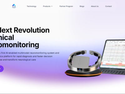
NeuroAstra is an AI-enabled EEG analysis and reporting software that provides a multimodal clinical EEG web platform with Co-Pilot AI to help neuro care teams detect abnormal trends faster and standardize reporting.
Cercare Medical Oncology Virtual Expert
Cercare Medical
Neurology AI
Cercare Medical's Oncology Virtual Expert is an AI-powered brain tumor segmentation module that provides fast, consistent, and semi-automated tumor analysis from standard MRI data, designed for on-premise deployment within existing clinical radiology workflows.
CoLumbo C-Spine
Smart Soft Healthcare AD
Clinical Decision Support & Reference
CoLumbo C-Spine is an AI-powered software that processes and analyzes cervical spine MRI exams, assisting clinicians in diagnosis and assessment.
MeRT System
Wave Neuroscience
Mental & Behavioral Health AI
MeRT (Magnetic e-Resonance Therapy) is an advanced, personalized form of TMS that uses your own brainwave data to guide treatment. It's a non-invasive, drug-free option designed to help the brain communicate more efficiently so symptoms of conditions like depression, anxiety, PTSD, TBI, autism, and more can improve.

See all Neurology AI tools →

More from Olea Medical

breastscape
Olea Medical
Oncology AI
breastscape by Olea Medical is an FDA-cleared AI-powered software for breast MRI post-processing, offering semi-automatic lesion diagnosis, BI-RADS reporting, and MRI-guided biopsy planning to enhance diagnostic confidence and streamline radiologist workflows.
MR DWI/FLAIR Measurement
Olea Medical
Neurology AI
Olea Medical's MR DWI/FLAIR Measurement V1.0 is an FDA-cleared AI-powered PACS software module for analyzing MR Diffusion-weighted imaging (DWI) and FLAIR images, providing critical metrics for neurological assessment and stroke diagnosis.

View company profile →

Suggest an Edit → | Report an issue | Last Verified: 2026-04-20 | First Added: 2026-04-20
AI Tool Finder
AI-powered search. Results may not be comprehensive.